
What should you know about pt3a melanoma?
Short answer
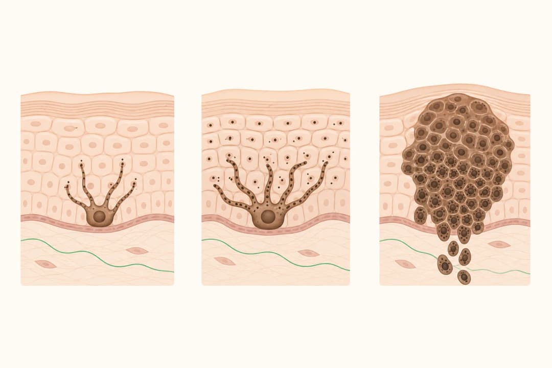
pT3a melanoma is an invasive melanoma in the pT3 thickness range without ulceration identified in the primary tumour.
This answers one question. It is not a diagnosis and does not replace an assessment by a licensed doctor.
At a glance
- pT3a
- A primary-tumour category based on a T3 thickness range without ulceration.
- Separate issue
- Lymph-node and distant findings are assessed outside the pT label.
- Care plan
- Excision and possible sentinel-node assessment are reviewed with a specialist.
Understanding the pathology
T3 describes the measured thickness of the primary melanoma, while a means the pathologist did not identify ulceration. Thickness is a microscope measurement, not an estimate based on how large the spot looks on the surface. The result describes the primary skin tumour only; it does not establish whether regional lymph nodes or distant organs contain melanoma.
Your report may mention margins, mitotic activity, and the biopsy method. These findings help specialists decide whether more tissue should be removed and whether additional staging is useful. Ask for a plain-language explanation of every term that affects your plan.
Treatment planning
A specialist will usually consider surgical excision of the biopsy site with a planned margin. The final specimen confirms whether the edges are free of melanoma. With a pT3 primary, sentinel lymph node biopsy is commonly discussed as a way to examine the first draining node pathway, but the decision depends on your circumstances and informed consent.
Take your pathology and operation details to each appointment. Follow wound-care instructions, use shade and protective clothing over healing skin, and begin regular self-checks once the team shows you what to look for.
When to seek care
Seek prompt review for a changing spot, a new pigmented lesion, a growing lump near the scar, or persistent bleeding. After treatment, contact the surgical team about fever, spreading redness, increasing warmth, discharge, wound opening, or pain that is becoming stronger. Emergency assessment is needed for sudden weakness, speech trouble, a seizure, marked confusion, or a severe new headache.
Questions for your consultation
Ask how thickness was measured, whether ulceration and margins were recorded, and what excision is recommended. Discuss the potential value and risks of sentinel lymph node biopsy, whether imaging is indicated, and how follow-up will monitor the scar, remaining skin, and lymph-node areas.
Find care
General Clinics & Polyclinics in Abu Dhabi
That answer is general. If you want it applied to your own case, these are licensed general clinics & polyclinics listed in Abu Dhabi.
27 Ar Ruwwad St - Shakhbout City - MSH4 - Abu Dhabi - United Arab Emirates
Accepts: Daman, Thiqa, Cigna, MetLife +20 more
Al Ruwais, Al Dhafrah
Accepts: Daman, Thiqa, AXA, Cigna +16 more
Al Bahyah - Al Bahyah Old - Abu Dhabi - United Arab Emirates
Accepts: Daman, Thiqa, Cigna, Allianz Care +18 more
Abu Dhabi
Accepts: Daman, Thiqa, AXA, Cigna +27 more
Ordered by verification status and how complete each clinic's public record is, then alphabetically. This is a directory listing, not a ranking — Zavis does not rate or recommend providers.
A factual listing drawn from the DHA, DOH and MOHAP licence registers. It is not a referral, an endorsement, or advice that any of these providers is right for you.
Questions people ask
Can the surface size predict the pT3 category?
No. The category uses microscopic thickness rather than the visible width of the lesion.
What happens after excision?
The specimen is examined for margins and other features, then the team discusses surveillance or further care.
Related
Related reading
Keep reading
Medical disclaimer. This page is general health information, not medical advice, and it cannot diagnose you. Always consult a licensed healthcare provider about your own symptoms and treatment. In an emergency in the UAE, call 998 for an ambulance.