
Pelvic MRI
At a glance
- Imaging method
- Magnetic fields and radio waves
- Radiation
- No ionising radiation
- Main focus
- Pelvic organs and nearby soft tissues
Find care
Radiology & Imaging in Dubai
Licensed radiology & imaging listed in Dubai. Appearing here means a provider holds a UAE licence in this category — not that it offers pelvic MRI, which is worth confirming with the clinic before you book.
First Gulf Real Estate Building, Amman Street, AL NAHDA FIRST, Dubai
Accepts: Daman, Thiqa, AXA, Cigna +28 more
LEADER, 3 Street 99, DUBAI INVESTMENT PARK FIRST, Dubai
Accepts: Daman, Thiqa, AXA, Cigna +29 more
villa, Al Wasl Road, JUMEIRAH FIRST, Dubai
Accepts: Daman, Thiqa, AXA, Cigna +16 more
Aletihad Diagnostic Center, Alwasl Road, AL SAFFA SECOND, Dubai
Accepts: AXA, Cigna, MetLife, Allianz Care +17 more
Moza Suwaidan Saeed Building, 45b Street 27, AL MURQABAT, Dubai
Accepts: Thiqa, AXA, Cigna, MetLife +25 more
Rdk1, 70 Street, DUBAI INVESTMENT PARK FIRST, Dubai
Accepts: Daman, AXA, Cigna, MetLife +20 more
Ordered by verification status and how complete each clinic's public record is, then alphabetically. This is a directory listing, not a ranking — Zavis does not rate or recommend providers.
A factual listing drawn from the DHA, DOH and MOHAP licence registers. It is not a referral, an endorsement, or advice that any of these providers is right for you.
What is a pelvic MRI?
A pelvic MRI creates detailed images of the uterus, ovaries, cervix, bladder, and nearby pelvic tissues without using X-rays.
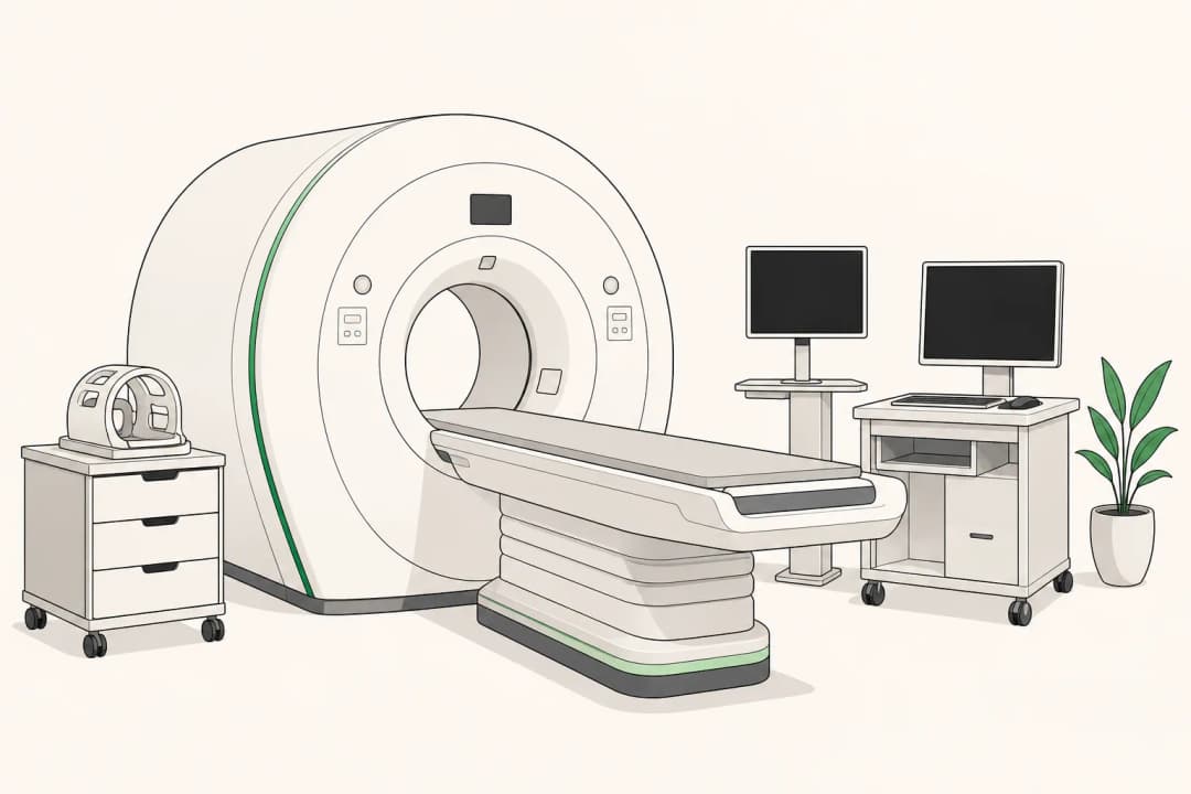
You lie on a motorised table that moves into a short, tunnel-shaped scanner. The scanner makes images from several angles, while a technologist monitors you and communicates through an intercom. The test can help clinicians examine soft tissues and map the size, position, and appearance of a finding.
What does the test involve?
Before the scan, you may be asked about metal implants, previous operations, kidney problems, allergies, and the possibility of pregnancy. You usually remove jewellery and other metal objects. Some examinations use contrast material through a vein to make certain tissues easier to distinguish; the imaging team will explain whether it is needed.
During the scan, staying still helps prevent blurred images. The machine makes repeated knocking or thumping sounds, so you may receive headphones or ear protection. You can usually speak with the technologist and use a call button if you feel uncomfortable.
Is it safe, and how accurate is it?
MRI does not use ionising radiation. Safety depends on the scanner, your medical history, and any implanted or retained metal. Tell the team about pacemakers, clips, shrapnel, medication pumps, or other implants before entering the scan room.
The images are detailed, but no scan answers every question. Movement, the body position, the area being examined, and whether contrast is appropriate can affect what is visible. A radiologist interprets the images alongside your symptoms, examination, and other test results.
What happens if the result is abnormal?
An abnormal result does not by itself identify the cause. It may describe a structural variation, inflammation, a cyst, a fibroid, or another area that needs closer assessment. Your clinician will explain the finding in context and may compare it with earlier images.
Next steps can include observation, a different scan, blood tests, a physical examination, or a tissue sample, depending on the appearance and your symptoms. For example, MRI can clarify the shape of the uterus when assessing a [Bicornuate Uterus](/conditions/bicornuate-uterus/).
Key facts about pelvic MRI
Pelvic MRI is commonly used when detailed soft-tissue information is needed or when another examination leaves an unanswered question. It can show relationships between organs and help with planning follow-up or treatment.
Who may need a pelvic MRI?
A clinician may request this scan for pelvic pain, unusual bleeding, a suspected mass, an unclear ultrasound finding, or a known condition that needs mapping. It may also help assess the uterus, ovaries, cervix, pelvic floor, bladder, or nearby lymph nodes.
The request should match the clinical question. Share your symptoms, past imaging, operations, pregnancies, and any implanted devices so the imaging team can select a safe and useful protocol.
How the scan is performed
The appointment usually follows these steps: change into suitable clothing; complete the safety screening; receive instructions about food, drink, or bladder preparation if relevant; lie on the padded table; allow the technologist to position a receiving device over the pelvis; and remain still while several image sequences are collected.
The table may move out briefly between sequences. If contrast is planned, it is given through a small intravenous line and the team watches for any reaction. The exact appointment length varies with the protocol and whether contrast is used.
After a pelvic MRI
Most people return to normal activities after a non-sedated MRI. You can usually eat, drink, and take regular medicines unless your care team gives different instructions. If contrast was used, follow the imaging centre's advice and tell them promptly about a rash, swelling, or breathing difficulty.
Possible risks and limitations
Some people feel confined, warm, or uncomfortable from lying still. Sedation may be discussed if claustrophobia is severe, but it requires extra planning and someone to take you home. Contrast can cause side effects, and kidney disease may affect whether it is suitable. Metal safety screening is essential.
Possible alternatives
Depending on the question, alternatives may include pelvic ultrasound, computed tomography, examination, blood tests, or follow-up imaging. Ultrasound can be useful for a quick assessment, while MRI may provide more detail about soft tissues. Your clinician chooses based on the suspected problem, urgency, pregnancy status, and safety factors.
Finding a pelvic MRI service
Choose an imaging service that can review your medical history, screen implants carefully, provide hearing protection, and explain contrast or sedation. Bring previous scan reports and tell the team what question your clinician wants answered. Ask when and how the report will be shared with the clinician managing your care.
Questions people ask
Can I have a pelvic MRI if I am pregnant?
Tell the imaging team if you are pregnant or might be pregnant. They will weigh the clinical need and choose the safest approach.
What if I am claustrophobic?
Tell the service when booking. The team can explain the scanner, offer communication during the test, and discuss preparation or sedation when appropriate.
Will I receive the result immediately?
A radiologist reviews the images and sends a report to the clinician who requested the scan. Timing depends on the service.
Related
Related reading
Keep reading
Medical disclaimer. This page is general health information, not medical advice, and it cannot diagnose you. Always consult a licensed healthcare provider about your own symptoms and treatment. In an emergency in the UAE, call 998 for an ambulance.