
Understanding the differential diagnosis of chondrosarcoma
Short answer
The differential diagnosis of chondrosarcoma compares it with benign cartilage tumours and other bone lesions that can look similar on scans or biopsy.
This answers one question. It is not a diagnosis and does not replace an assessment by a licensed doctor.
At a glance
- Key comparison
- Cancerous versus non-cancerous cartilage lesions
- Diagnosis combines
- Imaging, pathology and clinical history
- If findings disagree
- Specialist review or additional sampling may be considered
The short answer
The main distinction is often between a lower-grade chondrosarcoma and a non-cancerous cartilage lesion. Other possibilities depend on the tumour's site and appearance and can include different cartilage-producing or bone-forming tumours. No single symptom or scan phrase settles every case.
How specialists narrow the possibilities
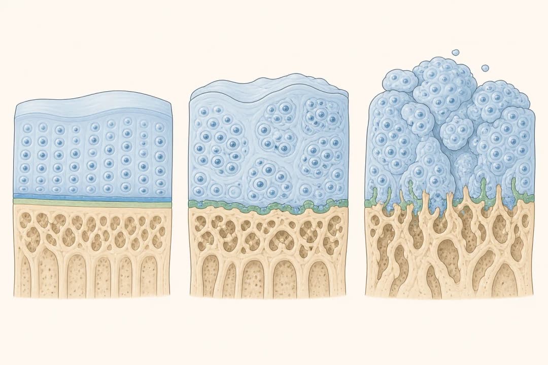
Radiologists assess the pattern of calcification, changes in the bone cortex, growth into soft tissue, tumour location and comparison with earlier images. Pathologists examine cell appearance, cartilage structure, growth into existing bone and any higher-grade or non-cartilaginous areas. Clinical details such as pain behaviour and documented growth provide additional context.
A small biopsy can miss a more active region within a varied tumour. For that reason, the biopsy route and target are planned from the imaging, and the pathology result is matched back to the sampled area. Disagreement between the scan and tissue findings may lead to slide review, repeat sampling or further imaging.
What to do when the diagnosis is uncertain
Arrange review through a multidisciplinary bone-tumour service before an operation or another invasive procedure. Bringing the actual scan files and pathology material allows radiologists, pathologists and surgeons to compare the same lesion. Ask whether the biopsy path could be removed during definitive surgery, because poorly placed sampling can complicate later tumour removal.
Until the team clarifies bone strength, follow advice about weight-bearing and activity. Record changes in pain, swelling or function and mention any previous cartilage lesion or skeletal condition. Treatment should follow the final integrated diagnosis rather than one isolated word from a report.
When to seek care sooner
Seek prompt assessment if pain is rapidly worsening, swelling enlarges, movement declines, or new numbness or weakness develops. Sudden severe pain, inability to bear weight or an obvious change in limb shape can indicate a fracture and warrants urgent care. Breathing difficulty or chest pain requires emergency evaluation. These warning signs matter because of possible structural or other complications, not because they prove a particular diagnosis.
Questions that clarify the diagnosis
Ask which diagnoses remain possible, which imaging features favour each one, and whether the biopsy sampled the most informative area. It is also reasonable to ask if the scan and pathology agree, whether specialist review is recommended, how bone strength affects activity, and what additional finding would change the proposed treatment.
Questions people ask
Can a scan alone diagnose chondrosarcoma?
Some imaging patterns can be strongly suggestive, but specialists interpret them with symptoms, examination and, when needed, carefully planned tissue sampling. The required approach depends on the lesion's site and level of concern.
Why can a cartilage tumour biopsy be difficult to interpret?
Benign cartilage lesions and lower-grade chondrosarcomas may share microscopic features. A tumour can also vary from one area to another, making comparison between the sampled spot and the imaging essential.
Related
Related reading
Keep reading
Cancer care in the UAE
Hospitals & Medical Centers in Abu Dhabi
Most UAE cancer care is delivered inside hospital services rather than by standalone oncology clinics, so these are licensed hospitals & medical centers listed in Abu Dhabi. Confirm the specific cancer service exists before you travel, and know that asking for a second opinion is routine.
Ahalia Hospital Musaffah Building - Musaffah - Musaffah Industrial - Abu Dhabi - United Arab Emirates
Accepts: Daman, Thiqa, AXA, Cigna +26 more
Al Ruwais Industrial City - Abu Dhabi - United Arab Emirates
Accepts: Daman, Thiqa, AXA, Cigna +30 more
Abu Dhabi, Abu Dhabi
Accepts: Daman, Thiqa, AXA, Cigna +30 more
Second opinions are routine in cancer care and asking for one does not affect your treatment. How to arrange a second opinion in the UAE.
Ordered by verification status and how complete each clinic's public record is, then alphabetically. This is a directory listing, not a ranking — Zavis does not rate or recommend providers.
A factual listing drawn from the DHA, DOH and MOHAP licence registers. It is not a referral, an endorsement, or advice that any of these providers is right for you.
Medical disclaimer. This page is general health information, not medical advice, and it cannot diagnose you. Always consult a licensed healthcare provider about your own symptoms and treatment. In an emergency in the UAE, call 998 for an ambulance.