
Lobar pneumonia on an X-ray
Short answer
A lobar pneumonia X-ray may show a dense, concentrated area of clouding in one lung lobe, but the image cannot identify every cause or predict severity alone.
This answers one question. It is not a diagnosis and does not replace an assessment by a licensed doctor.
At a glance
- Typical wording
- Lobar consolidation or focal clouding
- What it cannot do
- Identify every germ or measure severity by itself
- What guides care
- Symptoms, examination and oxygen level
How to understand the image
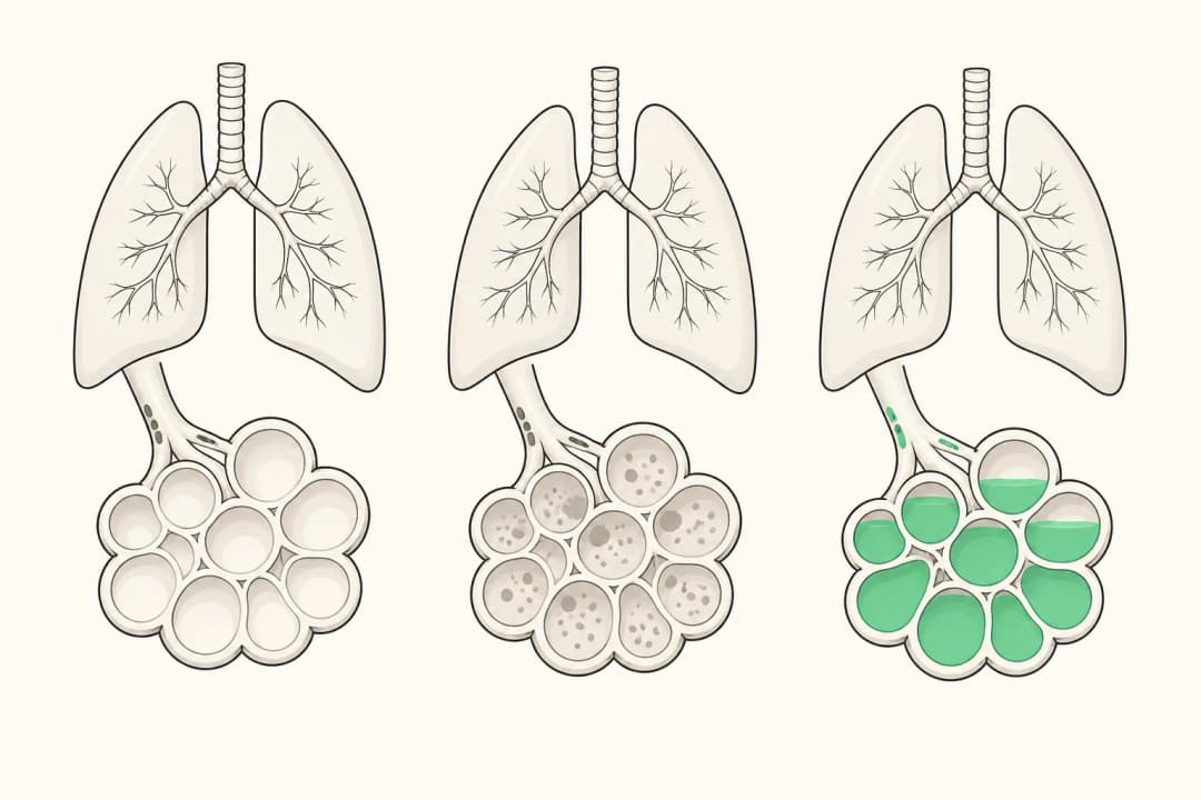
Healthy air spaces look darker on a chest X-ray because they contain air. Inflammation and fluid can make an affected region look whiter or denser. When that change follows the shape of a lobe, the report may use wording such as lobar consolidation. Similar appearances can occur with different infections and some non-infectious problems, so the report is interpreted alongside symptoms and examination.
A chest X-ray can support a diagnosis of pneumonia, but a normal or unclear image does not always settle the question early in an illness. Cough, fever, chills, phlegm and breathing difficulty remain relevant to the clinical assessment.
What happens after the X-ray
Your clinician may compare the X-ray with your symptoms, oxygen reading and lung examination. They may recommend monitoring, additional testing or treatment based on the likely cause and your overall health. Do not start or stop antibiotics because of the image alone. Follow the prescribed plan, drink fluids if you can, rest and report worsening breathlessness or new chest pain.
When to seek urgent care
Get urgent medical help for severe or quickly worsening breathlessness, blue or grey lips, confusion, fainting, severe chest pain or marked drowsiness. Contact a clinician promptly if fever continues, you cannot drink, or your cough and breathing are deteriorating. These changes matter even if an earlier X-ray result sounded limited.
Questions for your doctor
Ask what the report means in your case, whether the change is definitely in one lobe, what other causes were considered, whether repeat imaging is needed, and how you will know treatment is working.
Find care
General Clinics & Polyclinics in Abu Dhabi
That answer is general. If you want it applied to your own case, these are licensed general clinics & polyclinics listed in Abu Dhabi.
27 Ar Ruwwad St - Shakhbout City - MSH4 - Abu Dhabi - United Arab Emirates
Accepts: Daman, Thiqa, Cigna, MetLife +20 more
Al Ruwais, Al Dhafrah
Accepts: Daman, Thiqa, AXA, Cigna +16 more
Al Bahyah - Al Bahyah Old - Abu Dhabi - United Arab Emirates
Accepts: Daman, Thiqa, Cigna, Allianz Care +18 more
Abu Dhabi
Accepts: Daman, Thiqa, AXA, Cigna +27 more
Ordered by verification status and how complete each clinic's public record is, then alphabetically. This is a directory listing, not a ranking — Zavis does not rate or recommend providers.
A factual listing drawn from the DHA, DOH and MOHAP licence registers. It is not a referral, an endorsement, or advice that any of these providers is right for you.
Questions people ask
Can an X-ray prove which germ caused pneumonia?
No. The pattern can support pneumonia but usually cannot name the organism on its own.
Can pneumonia be present with an unclear X-ray?
Yes. Early or subtle illness may need clinical follow-up and, when appropriate, further assessment.
Medical disclaimer. This page is general health information, not medical advice, and it cannot diagnose you. Always consult a licensed healthcare provider about your own symptoms and treatment. In an emergency in the UAE, call 998 for an ambulance.