
Understanding an amelanotic melanoma pathology outline
Short answer
An amelanotic melanoma pathology outline explains how a pigment-poor melanoma was identified and records features that guide staging, treatment and follow-up.
This answers one question. It is not a diagnosis and does not replace an assessment by a licensed doctor.
At a glance
- Meaning of amelanotic
- The tumour has little or no visible melanin pigment
- Diagnostic approach
- Microscopic pattern, clinical context and a panel of special marker stains
- Key report details
- Thickness, ulceration, cell division, invasion and margin status when assessable
How pathologists recognise it
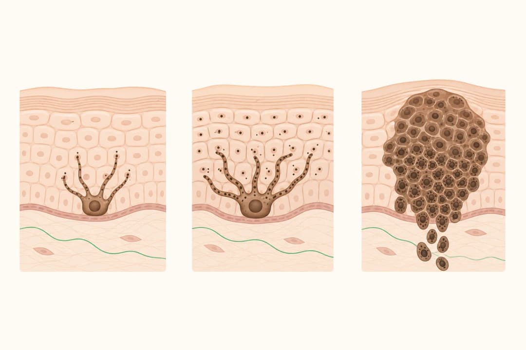
Amelanotic means that little or no melanin is visible in the tumour, so the lesion may not have the brown or black appearance associated with melanoma. Under the microscope, its cells can resemble other cancers. The pathologist evaluates the cell pattern and usually interprets special marker stains together rather than relying on colour or any single marker.
A final report may name the biopsy procedure and body site, confirm the diagnosis, and describe tumour thickness, ulceration, cell division, invasion into nearby structures and regression. If tissue was removed, margin findings indicate whether melanoma reaches an inked cut surface. A comment or diagnostic note may explain uncertainty or recommend correlation with the clinical findings.
How to use the report
Review the pathology result with the clinician who performed the biopsy. Confirm whether the sample removed the whole lesion or only part of it, because a partial biopsy may not show the deepest point. Ask which findings determine the pathological stage and whether the margins can be assessed from this specimen.
Next steps may include wider excision, assessment of regional lymph nodes, imaging or an oncology consultation. The choice depends on the complete pathology report, examination findings and your overall health. Keep a copy of the original report and any later excision report so that the team can compare the diagnosis, depth and margins.
When to contact the care team promptly
Contact the treating clinic promptly if a biopsy wound has worsening redness, swelling, discharge, persistent bleeding or increasing pain. Also report a rapidly changing lesion, a new lump near the affected area or unexplained neurological symptoms. Seek emergency care for severe bleeding, sudden weakness, a seizure, new confusion or difficulty breathing, whatever the presumed cause.
Questions to ask about the findings
Ask what supported the melanoma diagnosis despite the absent pigment, whether the sample was deep enough for measurement, and which features affect staging. Clarify whether melanoma is present at a margin, whether another procedure is advised, whether lymph-node assessment would add useful information and who will coordinate skin surveillance after treatment.
Find care
General Clinics & Polyclinics in Abu Dhabi
That answer is general. If you want it applied to your own case, these are licensed general clinics & polyclinics listed in Abu Dhabi.
27 Ar Ruwwad St - Shakhbout City - MSH4 - Abu Dhabi - United Arab Emirates
Accepts: Daman, Thiqa, Cigna, MetLife +20 more
Al Ruwais, Al Dhafrah
Accepts: Daman, Thiqa, AXA, Cigna +16 more
Al Bahyah - Al Bahyah Old - Abu Dhabi - United Arab Emirates
Accepts: Daman, Thiqa, Cigna, Allianz Care +18 more
Abu Dhabi
Accepts: Daman, Thiqa, AXA, Cigna +27 more
Ordered by verification status and how complete each clinic's public record is, then alphabetically. This is a directory listing, not a ranking — Zavis does not rate or recommend providers.
A factual listing drawn from the DHA, DOH and MOHAP licence registers. It is not a referral, an endorsement, or advice that any of these providers is right for you.
Questions people ask
Why are special stains used for amelanotic melanoma?
Without obvious melanin, the tumour may resemble another type of cancer under routine microscopy. Marker stains help the pathologist establish the cell lineage, with the pattern interpreted alongside the tissue appearance and clinical site.
What does a positive margin mean?
It means melanoma cells extend to a cut edge of the submitted tissue. The treating team uses that finding with the procedure type and lesion site to decide whether further local treatment should be discussed.
Can a partial biopsy provide the final tumour thickness?
Not always. If the biopsy does not include the deepest part of the lesion, the pathologist may be unable to give a definitive thickness. A later excision can provide additional information.
Related
Related reading
Keep reading
Medical disclaimer. This page is general health information, not medical advice, and it cannot diagnose you. Always consult a licensed healthcare provider about your own symptoms and treatment. In an emergency in the UAE, call 998 for an ambulance.