
What should you know about pt1a melanoma prognosis?
Short answer
Pt1a melanoma is an early-stage melanoma with a generally favourable outlook, but your personal prognosis depends on the pathology report, treatment and follow-up findings.
This answers one question. It is not a diagnosis and does not replace an assessment by a licensed doctor.
At a glance
- Stage label
- pT1a is a pathological stage for a thin, non-ulcerated primary melanoma.
- Main prognostic clues
- Tumour depth, ulceration, margins, examination findings and any evidence of spread.
- Follow-up focus
- The scar, nearby lymph nodes and the rest of the skin.
The short answer
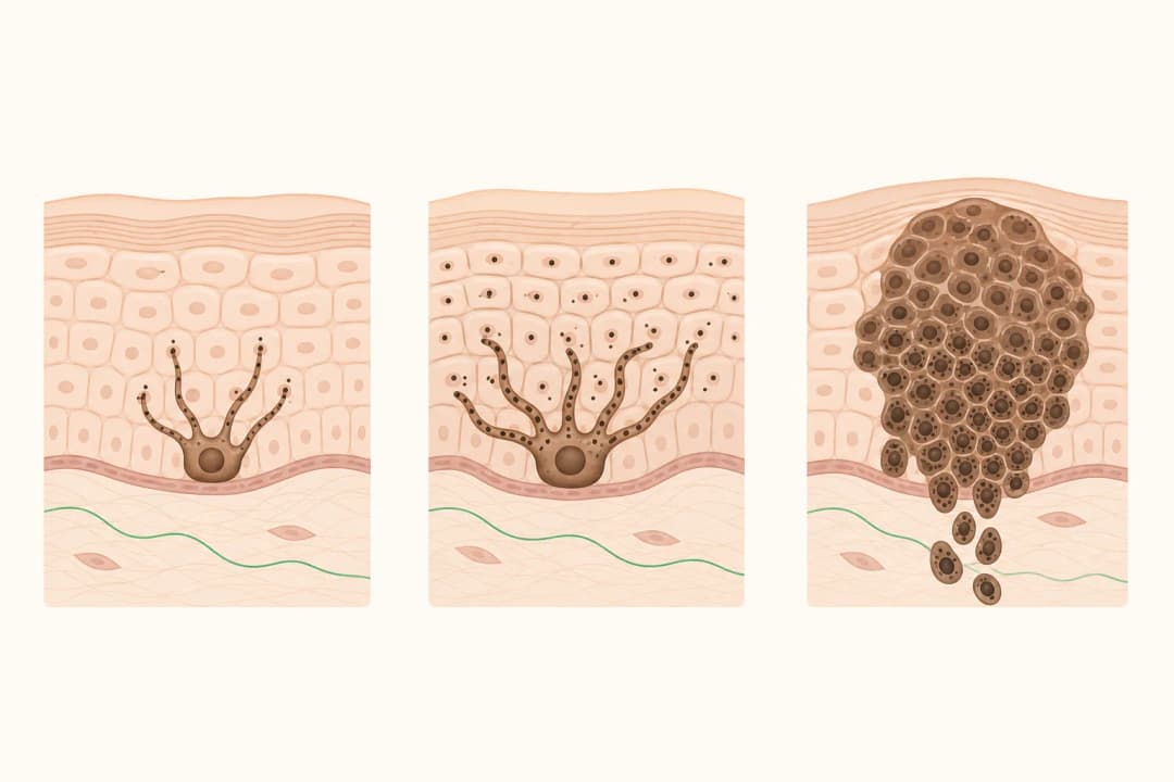
Pt1a melanoma is an early-stage melanoma with a generally favourable outlook, but your personal prognosis depends on the pathology report, treatment and follow-up findings. The label describes a thin primary melanoma without ulceration. It does not by itself predict exactly what will happen to one person. Your dermatology or oncology team combines the stage with the tumour's measured depth, examination findings and whether any additional testing is needed.
What pt1a means
The p in pT1a refers to a pathological assessment, meaning the stage is based on tissue examined by a pathologist. T1 describes a melanoma in the thinnest primary-tumour category. The a subdivision indicates that ulceration was not seen. The report may also mention mitotic activity, margins and other microscopic details; these findings help your clinician decide whether the initial removal was sufficient and how closely to monitor you.
A pt1a result is not the same as a diagnosis of spread. Lymph nodes, skin near the original site and distant organs are assessed separately when clinically indicated. A scan or procedure is not automatically needed for every person with a thin melanoma. A [Melanoma](/conditions/melanoma/) overview can help explain the disease and its staging terms.
What usually happens next
First, review the pathology report with the clinician who removed or diagnosed the lesion. Confirm the tumour depth, ulceration status, margin status and recorded stage. If more tissue needs to be removed around the original site, the procedure is called a wider local excision; read about [Surgical Excision](/treatments/surgical-dermatology/excisional-surgery/) and [Skin Cancer Surgery](/treatments/surgical-dermatology/skin-cancer-surgery/).
Follow-up commonly includes skin examinations and checking the area around the scar and nearby lymph nodes. Learn how [Follow-Up After Cancer Treatment](/treatments/survivorship-and-rehabilitation/post-treatment-surveillance/) may be organised. Protecting your skin from ultraviolet exposure and becoming familiar with your own moles can make new or changing marks easier to notice. Do not examine the scar so often that normal healing changes are mistaken for recurrence.
When to contact your care team
Contact your dermatology team promptly if the scar develops a new lump, persistent bleeding, a non-healing area or a firm change that does not settle. Arrange assessment for a new or changing mole, especially one with evolving colour, shape or size. A new swollen lymph node near the treated area also deserves clinical review.
Seek urgent medical attention for sudden neurological symptoms such as new weakness, confusion, a seizure or a severe new headache. These symptoms have many possible causes, but they should not be monitored at home. Information about [Brain Metastases](/conditions/brain-metastases/) explains why neurological changes are assessed carefully in people with a history of cancer.
Questions for your doctor
Ask: What are the measured depth and margin findings in my report? Is wider excision recommended? Do my examination findings suggest any need for a [Sentinel Lymph Node Biopsy](/treatments/diagnostics-and-staging/sentinel-lymph-node-biopsy/)? How often should my skin and lymph nodes be checked? Which changes should prompt an earlier visit? If new findings change the stage, ask whether [Immunotherapy](/treatments/systemic-therapy/immunotherapy/) or another treatment is relevant.
Find care
General Clinics & Polyclinics in Abu Dhabi
That answer is general. If you want it applied to your own case, these are licensed general clinics & polyclinics listed in Abu Dhabi.
27 Ar Ruwwad St - Shakhbout City - MSH4 - Abu Dhabi - United Arab Emirates
Accepts: Daman, Thiqa, Cigna, MetLife +20 more
Al Ruwais, Al Dhafrah
Accepts: Daman, Thiqa, AXA, Cigna +16 more
Al Bahyah - Al Bahyah Old - Abu Dhabi - United Arab Emirates
Accepts: Daman, Thiqa, Cigna, Allianz Care +18 more
Abu Dhabi
Accepts: Daman, Thiqa, AXA, Cigna +27 more
Ordered by verification status and how complete each clinic's public record is, then alphabetically. This is a directory listing, not a ranking — Zavis does not rate or recommend providers.
A factual listing drawn from the DHA, DOH and MOHAP licence registers. It is not a referral, an endorsement, or advice that any of these providers is right for you.
Questions people ask
Does pt1a melanoma mean the cancer has spread?
No. Pt1a describes the primary melanoma's thinness and lack of ulceration. Spread is assessed separately through examination and further tests when there is a clinical reason.
Will pt1a melanoma need immunotherapy?
Many people with an early, fully removed melanoma do not need systemic treatment. Your team considers treatment if additional findings change the stage or indicate a higher risk.
Can pt1a melanoma come back?
Recurrence is possible, so scheduled skin checks and attention to new or changing lesions remain useful even after treatment of an early melanoma.
Related
Related reading
Keep reading
Medical disclaimer. This page is general health information, not medical advice, and it cannot diagnose you. Always consult a licensed healthcare provider about your own symptoms and treatment. In an emergency in the UAE, call 998 for an ambulance.