
Melanoma staging: reading a pathology outline
Short answer
A melanoma pathology outline records tumour features such as thickness, ulceration, margins, and node findings that help determine the stage.
This answers one question. It is not a diagnosis and does not replace an assessment by a licensed doctor.
At a glance
- Core measurement
- Tumour thickness
- Surface feature
- Ulceration
- Surgical detail
- Margin status
Terms you may see
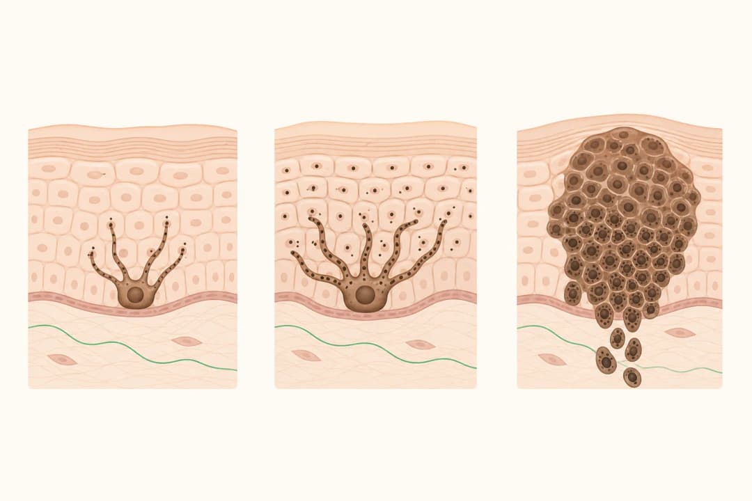
The report identifies the melanoma type and describes how deeply it extends into the skin. Thickness is measured in millimetres and is interpreted with other findings. Ulceration means the surface over the tumour is absent; its presence can affect staging. The report also describes whether the edges, or margins, contain melanoma cells and may comment on mitotic activity or microsatellites.
If a lymph node was sampled, the report states whether melanoma cells were found there and may describe the amount and location of involvement. These words are pieces of a clinical picture, not a diagnosis to interpret in isolation. Read more about [Melanoma] and the role of [Sentinel Lymph Node Biopsy].
How to use the report
Ask your dermatologist or surgical oncologist to mark the exact lines that determine stage and explain any terms that remain unclear. Confirm whether the excision removed the lesion with clear margins and whether more surgery is advised. [Surgical Excision] is commonly used for removal of the primary melanoma, while [Skin Cancer Surgery] covers the wider surgical context.
Keep the report with your medical records and bring it to appointments. Do not compare a single thickness measurement with another person’s case: staging also considers ulceration, nodes, distant findings, and the quality of the available examination.
When to seek care
Contact the clinic if you cannot access the pathology report, if the planned results appointment has not been arranged, or if the wound develops spreading redness, discharge, increasing pain, or fever. Report a new lump near the scar or in the draining lymph-node area. Persistent neurological symptoms such as new weakness, confusion, or severe headache also warrant prompt medical assessment.
Questions for your doctor
Which words in my report determine the stage? What is the tumour thickness and is ulceration present? Are the margins clear? Was a lymph node examined, and what did it show? Is another procedure or scan needed to complete staging?
Find care
General Clinics & Polyclinics in Abu Dhabi
That answer is general. If you want it applied to your own case, these are licensed general clinics & polyclinics listed in Abu Dhabi.
27 Ar Ruwwad St - Shakhbout City - MSH4 - Abu Dhabi - United Arab Emirates
Accepts: Daman, Thiqa, Cigna, MetLife +20 more
Al Ruwais, Al Dhafrah
Accepts: Daman, Thiqa, AXA, Cigna +16 more
Al Bahyah - Al Bahyah Old - Abu Dhabi - United Arab Emirates
Accepts: Daman, Thiqa, Cigna, Allianz Care +18 more
Abu Dhabi
Accepts: Daman, Thiqa, AXA, Cigna +27 more
Ordered by verification status and how complete each clinic's public record is, then alphabetically. This is a directory listing, not a ranking — Zavis does not rate or recommend providers.
A factual listing drawn from the DHA, DOH and MOHAP licence registers. It is not a referral, an endorsement, or advice that any of these providers is right for you.
Questions people ask
Does a pathology outline alone tell the whole story?
It provides essential tumour information, but final staging may also use examination, lymph-node findings, and selected imaging.
What does a clear margin mean?
It means no melanoma cells were identified at the examined edge of the removed tissue; your specialist explains whether further treatment is needed.
Related
Related reading
Keep reading
Medical disclaimer. This page is general health information, not medical advice, and it cannot diagnose you. Always consult a licensed healthcare provider about your own symptoms and treatment. In an emergency in the UAE, call 998 for an ambulance.