
What should you know about lentigo maligna melanoma pathology outlines?
Short answer
A pathology outline for lentigo maligna melanoma describes the abnormal cells, invasion, margins, and features that guide staging and treatment decisions.
This answers one question. It is not a diagnosis and does not replace an assessment by a licensed doctor.
At a glance
- Confirms diagnosis
- Microscopic examination of tissue.
- Key planning details
- Invasion, thickness, ulceration, and specimen margins.
What the report examines
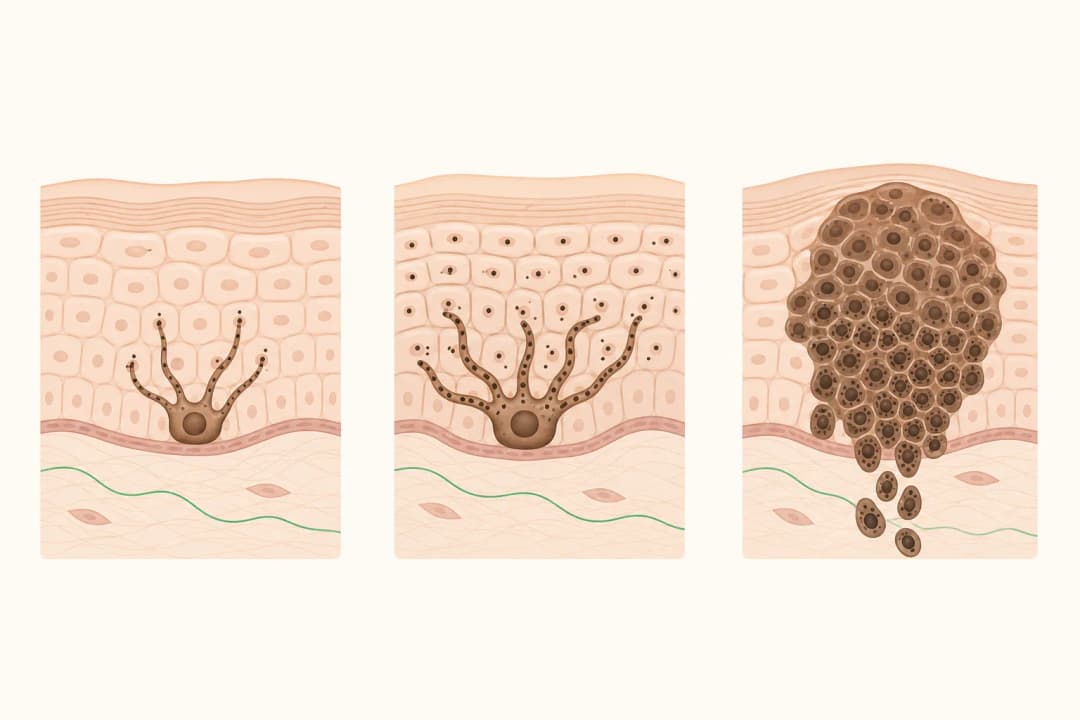
The pathologist studies the biopsy under a microscope to determine whether melanoma is present and whether it is limited to the outer skin or has invaded deeper tissue. The report may describe tumour thickness, ulceration, cell division, and whether the edges of the specimen are clear. These details help the treating team estimate the extent of disease and plan the next step.
A report can contain unfamiliar terms and does not, on its own, predict exactly what will happen to one person. A specialist may review the slides or request additional testing when the result is difficult to interpret.
What to do after the result
Ask for a copy of the report and arrange a consultation to discuss the diagnosis, margin status, and whether more tissue should be removed. Treatment may involve surgical excision; some people need assessment of nearby lymph nodes or scans depending on the findings. Keep the biopsy site clean as instructed and record new skin changes for follow-up.
When to seek care
Contact the clinical team promptly if the wound keeps bleeding, opens, becomes increasingly painful, or develops spreading redness, warmth, drainage, or fever. Before the planned review, report a new lump near the biopsy site, swollen lymph nodes, persistent headaches, weakness, seizures, or changes in vision. These symptoms need clinical assessment and should not be interpreted from the pathology wording alone.
Questions for your doctor
Ask whether the sample shows melanoma or an early non-invasive form, whether the margins are clear, and whether the reported depth changes management. Ask if further excision, lymph-node assessment, imaging, or oncology input is appropriate.
Find care
General Clinics & Polyclinics in Abu Dhabi
That answer is general. If you want it applied to your own case, these are licensed general clinics & polyclinics listed in Abu Dhabi.
27 Ar Ruwwad St - Shakhbout City - MSH4 - Abu Dhabi - United Arab Emirates
Accepts: Daman, Thiqa, Cigna, MetLife +20 more
Al Ruwais, Al Dhafrah
Accepts: Daman, Thiqa, AXA, Cigna +16 more
Al Bahyah - Al Bahyah Old - Abu Dhabi - United Arab Emirates
Accepts: Daman, Thiqa, Cigna, Allianz Care +18 more
Abu Dhabi
Accepts: Daman, Thiqa, AXA, Cigna +27 more
Ordered by verification status and how complete each clinic's public record is, then alphabetically. This is a directory listing, not a ranking — Zavis does not rate or recommend providers.
A factual listing drawn from the DHA, DOH and MOHAP licence registers. It is not a referral, an endorsement, or advice that any of these providers is right for you.
Questions people ask
What does a clear margin mean?
It means no melanoma cells were seen at the examined edge of that specimen, although the care team decides whether further treatment is needed.
Why might more surgery be recommended?
A wider excision may remove surrounding tissue when the first sample does not provide the planned safety margin or when invasive disease is present.
Related
Related reading
Keep reading
Medical disclaimer. This page is general health information, not medical advice, and it cannot diagnose you. Always consult a licensed healthcare provider about your own symptoms and treatment. In an emergency in the UAE, call 998 for an ambulance.