
Amelanotic nodular melanoma explained
Short answer
Amelanotic nodular melanoma is a raised, often fast-growing melanoma with little visible pigment, so a persistent pink, red or skin-coloured nodule needs prompt clinical assessment.
This answers one question. It is not a diagnosis and does not replace an assessment by a licensed doctor.
At a glance
- Typical form
- A raised or dome-shaped nodule
- Possible colour
- Pink, red or skin-coloured, sometimes with a darker area
- Confirmation
- A biopsy is needed for diagnosis
The key point
It may resemble a harmless bump, inflamed spot or sore because it lacks the brown or black colour many people associate with melanoma. Appearance alone cannot confirm it; diagnosis requires a clinician's examination and a biopsy.
What the name describes
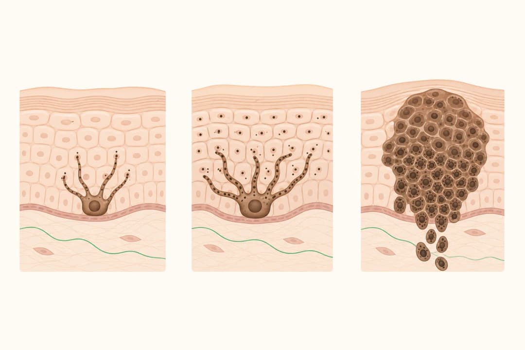
“Amelanotic” means the lesion has little or no obvious melanin pigment. “Nodular” describes a growth pattern that tends to form a firm, dome-shaped or raised nodule and may grow down into the skin. Some lesions retain a small darker area, while others appear uniformly pink or red.
Possible surface changes include bleeding, crusting, tenderness or ulceration, although a lesion may have none of these. Nodular melanoma can arise on otherwise normal-looking skin. A clinician may use magnification to inspect it, but tissue examination is needed to distinguish it from other skin growths.
What to do about a suspicious nodule
Book a skin assessment without delay if a raised growth is enlarging, repeatedly bleeding or failing to heal. Record when you noticed it and take a clear photograph with a size reference if this does not postpone care. Mention changes in height as well as width and colour.
Avoid picking, shaving over or treating the nodule with acids, freezing products or wart remedies. These measures may cause bleeding and obscure its surface. If a biopsy confirms melanoma, the team uses the pathology details and staging assessment to plan surgical removal and decide whether lymph-node evaluation or systemic treatment should be considered.
Changes that need faster help
Seek prompt medical review for rapid growth, recurrent bleeding, a sore that stays open, new crusting or a firm nodule that keeps changing. These are reasons for timely assessment regardless of the lesion's colour or whether it hurts.
After a melanoma diagnosis, tell the treating team about new swelling or a lump near the lesion or in the draining lymph-node area. Emergency evaluation is appropriate for sudden severe breathlessness, collapse, a seizure, new one-sided weakness or abrupt confusion.
Useful questions at the consultation
You can ask which part of the lesion will be sampled, when and how results will be shared, and what wound care the biopsy site needs. If melanoma is confirmed, ask about its thickness and ulceration, whether the margins are clear, which staging steps are relevant, and what the proposed treatment is intended to achieve.
Find care
General Clinics & Polyclinics in Abu Dhabi
That answer is general. If you want it applied to your own case, these are licensed general clinics & polyclinics listed in Abu Dhabi.
27 Ar Ruwwad St - Shakhbout City - MSH4 - Abu Dhabi - United Arab Emirates
Accepts: Daman, Thiqa, Cigna, MetLife +20 more
Al Ruwais, Al Dhafrah
Accepts: Daman, Thiqa, AXA, Cigna +16 more
Al Bahyah - Al Bahyah Old - Abu Dhabi - United Arab Emirates
Accepts: Daman, Thiqa, Cigna, Allianz Care +18 more
Abu Dhabi
Accepts: Daman, Thiqa, AXA, Cigna +27 more
Ordered by verification status and how complete each clinic's public record is, then alphabetically. This is a directory listing, not a ranking — Zavis does not rate or recommend providers.
A factual listing drawn from the DHA, DOH and MOHAP licence registers. It is not a referral, an endorsement, or advice that any of these providers is right for you.
Questions people ask
Can amelanotic nodular melanoma look like a pimple?
It can resemble an inflamed bump or sore, particularly when it is pink or red. A nodule that persists, grows, bleeds or crusts should be examined rather than assumed to be acne.
Can it occur without a pre-existing mole?
Yes. Nodular melanoma may develop on skin where no mole was previously noticed, so change in any persistent new growth matters.
How is the diagnosis confirmed?
A clinician removes all or part of the suspicious lesion for examination. The pathology report identifies the tumour and provides features used for staging and treatment planning.
Related
Related reading
Keep reading
Medical disclaimer. This page is general health information, not medical advice, and it cannot diagnose you. Always consult a licensed healthcare provider about your own symptoms and treatment. In an emergency in the UAE, call 998 for an ambulance.